Beranda
/ Chest Muscles Anatomy - Chest Anatomy Muscle - Cheat Dumper - This video covers the definition, innervation and functions of the two pectoral muscles:
Chest Muscles Anatomy - Chest Anatomy Muscle - Cheat Dumper - This video covers the definition, innervation and functions of the two pectoral muscles:
Insurance Gas/Electricity Loans Mortgage Attorney Lawyer Donate Conference Call Degree Credit Treatment Software Classes Recovery Trading Rehab Hosting Transfer Cord Blood Claim compensation mesothelioma mesothelioma attorney Houston car accident lawyer moreno valley can you sue a doctor for wrong diagnosis doctorate in security top online doctoral programs in business educational leadership doctoral programs online car accident doctor atlanta car accident doctor atlanta accident attorney rancho Cucamonga truck accident attorney san Antonio ONLINE BUSINESS DEGREE PROGRAMS ACCREDITED online accredited psychology degree masters degree in human resources online public administration masters degree online bitcoin merchant account bitcoin merchant services compare car insurance auto insurance troy mi seo explanation digital marketing degree floridaseo company fitness showrooms stamfordct how to work more efficiently seowordpress tips meaning of seo what is an seo what does an seo do what seo stands for best seotips google seo advice seo steps, The secure cloud-based platform for smart service delivery. Safelink is used by legal, professional and financial services to protect sensitive information, accelerate business processes and increase productivity. Use Safelink to collaborate securely with clients, colleagues and external parties. Safelink has a menu of workspace types with advanced features for dispute resolution, running deals and customised client portal creation. All data is encrypted (at rest and in transit and you retain your own encryption keys. Our titan security framework ensures your data is secure and you even have the option to choose your own data location from Channel Islands, London (UK), Dublin (EU), Australia.
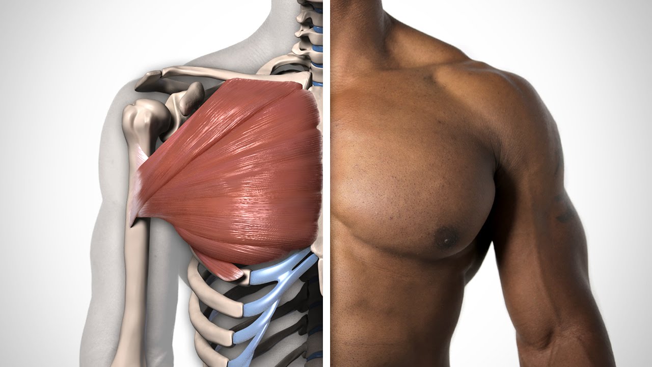
Chest Muscles Anatomy - Chest Anatomy Muscle - Cheat Dumper - This video covers the definition, innervation and functions of the two pectoral muscles:. Often referred to as the torso muscles or simply 'pecs', these thick muscles connect your chest to your upper arm and are responsible for a number of movements. Basically, the pectoralis minor is located directly underneath the pectoralis major. This video covers the definition, innervation and functions of the two pectoral muscles: Visceral muscles are found inside organs such as the stomach, intestines and blood vessels. A heart attack results from blocked blood flow, often from a blood clot, to your heart muscle.
Visceral muscles are found inside organs such as the stomach, intestines and blood vessels. Muscle anatomy crossword answer key Learn chest muscles human anatomy with free interactive flashcards. The pectoralis major, the larger and more superficial, originates at the clavicle (collarbone), the sternum, the ribs, and a tendinous extension. Overall, these chest muscles start at the clavicle and insert at the sternum and the armpit area (humerous).
How to Draw Pecs - Anatomy - YouTube from i.ytimg.com (1) the pectoralis major, and (2) the pectoralis minor. Angina is the term for chest pain caused by poor blood flow to the heart. Building chest muscles yields more than a chiseled chest. Chest muscles anatomy the chest is made up primarily of two muscles: Cardiac muscle is found in the heart. These important muscles control many motions that involve moving the arms and head — such as throwing a ball, looking up at the sky, and raising your hand. The muscles of the chest and upper back occupy the thoracic region of the body inferior to the neck and superior to the abdominal region and include the muscles of the shoulders. Moreover, the other muscular systems found inside the chest are digestive, circulatory and respiratory muscles which are not very noticeable, but they significantly have impact on complete human organism.
Learn about each of these muscles, their locations, functional anatomy and exercises for them.
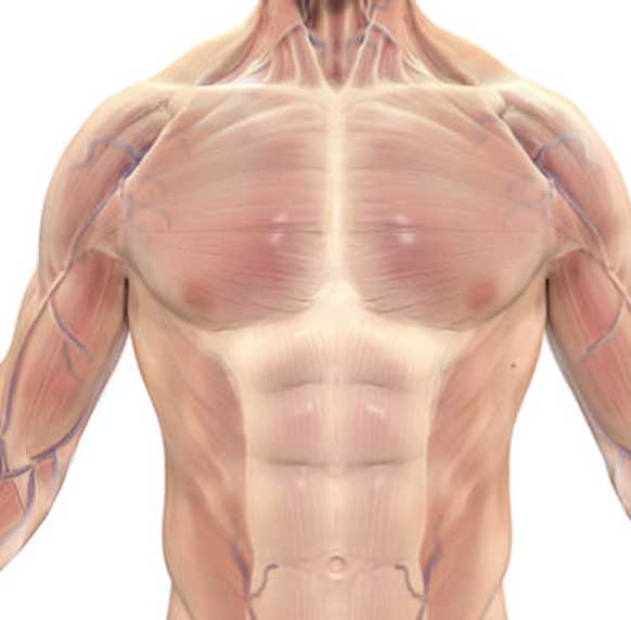
Chest pain has many possible causes, all of which need medical attention. Chest muscles anatomy the chest is made up primarily of two muscles: Here, we break down the anatomy of your chest muscles. Skeletal muscles are connected directly to bones by tendons (elastic type fibers). Cardiac muscle is found in the heart. It contains four muscles that exert a force on the upper limb: What you refer to as your chest is actually a group of called the pectorals. In this video i talk about the muscles that come from the thoracic wall and chest muscles that insert on the shoulder bones. Related posts of chest muscles diagram arm muscle anatomy diagram. All about the chest muscles the chest anatomy includes the pectoralis major, pectoralis minor and the serratus anterior. However, our primary focus is on the chest's anatomy or the chest's main muscles in this section. This page provides an overview of the chest muscle group. Choose from 500 different sets of chest muscles human anatomy flashcards on quizlet.
However, our primary focus is on the chest's anatomy or the chest's main muscles in this section. The pectoralis major, the larger and more superficial, originates at the clavicle (collarbone), the sternum, the ribs, and a tendinous extension. Pectoralis muscle, any of the muscles that connect the front walls of the chest with the bones of the upper arm and shoulder.there are two such muscles on each side of the sternum (breastbone) in the human body: The muscles of the chest and upper back occupy the thoracic region of the body inferior to the neck and superior to the abdominal region and include the muscles of the shoulders. Learn about each of these muscles, their locations, functional anatomy and exercises for them.
10+ Best Chest Exercises to Build INSANE Muscles: Complete ... from totalshape.com These important muscles control many motions that involve moving the arms and head — such as throwing a ball, looking up at the sky, and raising your hand. Here, we break down the anatomy of your chest muscles. See chest anatomy stock video clips. Computed tomography (ct) of the chest can detect pathology that may not show up on a conventional chest radiograph(1). Strength training exercises for the chest protect against diabetes and help you retain muscle mass during aging and weight loss. What you refer to as your chest is actually a group of called the pectorals. This mri chest (thorax) axial cross sectional anatomy tool is absolutely free to use. The muscles of the thoracic cage are the pectoralis major, pectoralis minor, serratus anterior, subclavius, intercostal (external, internal and innermost), subcostal and transversus thoracis muscles, including the diaphragm.
All about the chest muscles the chest anatomy includes the pectoralis major, pectoralis minor and the serratus anterior.
Learn about each of these muscles, their locations, functional anatomy and exercises for them. The chest's anatomy may explain why it gets sore and tight. The muscles of the chest and upper back occupy the thoracic region of the body inferior to the neck and superior to the abdominal region and include the muscles of the shoulders. A heart attack results from blocked blood flow, often from a blood clot, to your heart muscle. All about the chest muscles the chest anatomy includes the pectoralis major, pectoralis minor and the serratus anterior. Choose from 500 different sets of chest muscles human anatomy flashcards on quizlet. However, our primary focus is on the chest's anatomy or the chest's main muscles in this section. The terms pulled muscle and muscle strain refer to an injury that involves an overstretched or torn muscle. A person with a muscle strain in the chest may experience sudden, sharp pain in this area. The pectoralis major and the pectoralis minor, known collectively as your pecs. See chest anatomy stock video clips. It contains four muscles that exert a force on the upper limb: Chest muscles anatomy the chest is made up primarily of two muscles:
Related posts of chest muscles diagram arm muscle anatomy diagram. Clavicular head & sternocostal head. Lateral lip of intertubercular groove of the. Often referred to as the torso muscles or simply 'pecs', these thick muscles connect your chest to your upper arm and are responsible for a number of movements. A heart attack results from blocked blood flow, often from a blood clot, to your heart muscle.
Anatomy Of Stomach Muscles - Human Anatomy Diagram ... from i.pinimg.com The chest, or scientifically termed thorax, is located between the neck and abdomen, containing the thorax cavity and thorax wall. This mri chest (thorax) axial cross sectional anatomy tool is absolutely free to use. Building chest muscles yields more than a chiseled chest. Plus, how to target each to make them bigger and stronger. This page provides an overview of the chest muscle group. It's made up of the pectoralis major and pectoralis minor. Angina is the term for chest pain caused by poor blood flow to the heart. Related posts of chest muscles diagram arm muscle anatomy diagram.
Muscles of the chest and their functions you have two mighty muscles on both sides of your chest:
The muscles of the chest and upper back occupy the thoracic region of the body inferior to the neck and superior to the abdominal region and include the muscles of the shoulders. Chest muscles anatomy the chest is made up primarily of two muscles: Clavicular head & sternocostal head. Computed tomography (ct) of the chest can detect pathology that may not show up on a conventional chest radiograph(1). Arm muscle anatomy diagram 12 photos of the arm muscle anatomy diagram arm muscle anatomy diagram, human anatomy arm muscle diagram, human muscles, arm muscle anatomy diagram, human anatomy arm muscle diagram. Pectoralis major and pectoralis minor. Muscle anatomy crossword answer key Four main muscles in the pectoral region exert a force on the upper limb. Muscles the dominant muscle in the upper chest is the pectoralis major. The muscle are the pectoralis major and pectoralis minor. The pectoralis major, pectoralis minor, serratus anterior and subclavius. Use the mouse scroll wheel to move the images up and down alternatively use the tiny arrows (>>) on both side of the image to move the images.>>) on both side of the image to move the images. The chest's anatomy may explain why it gets sore and tight.